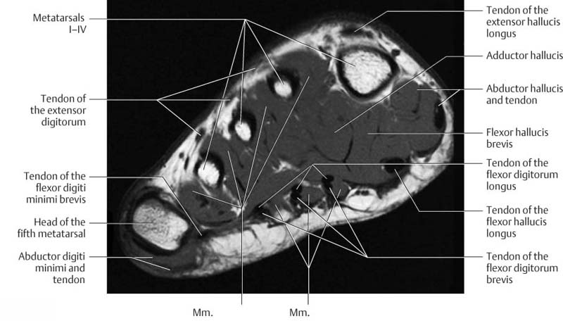
Foot Muscle Anatomy Coronal Mri. A wide array of supernumerary and accessory musculature has been described in the anatomic surgical and radiology literature. Lumbar spine anatomy on MRI Magnetic Resonance Imaging Anatomy of the lumbar spine using cross-sectional imaging MR T1 and T2 weighted.

Magnetic resonance imaging MRI is arguably the most sophisticated imaging method used in clinical medicineIn recent years MRI scans have become increasingly common as costs decrease. Lumbar spine anatomy on MRI Magnetic Resonance Imaging Anatomy of the lumbar spine using cross-sectional imaging MR T1 and T2 weighted. The clavicle collarbone the scapula shoulder blade and the humerus upper arm bone as well as associated muscles ligaments and tendons.
Spin-echo T1 and proton-density with fat saturation sequences.
In some cases however accessory muscles may produce clinical symptoms. The second peroneal tendon is found to have limited excursion with multiple tears and fibrous tissue. Magnetic resonance imaging MRI is arguably the most sophisticated imaging method used in clinical medicineIn recent years MRI scans have become increasingly common as costs decrease. Fat depositions are the result of chronic bowel inflammation and therefore quite common in.
