Hip Bone Anatomy Ct Scan. This CT imaging of the temporal bone was performed on a normal subject. 25-7 Experienced hip arthroscopists have reported low.
Normal Pelvis Radiology Case Radiopaedia Org from radiopaedia.org
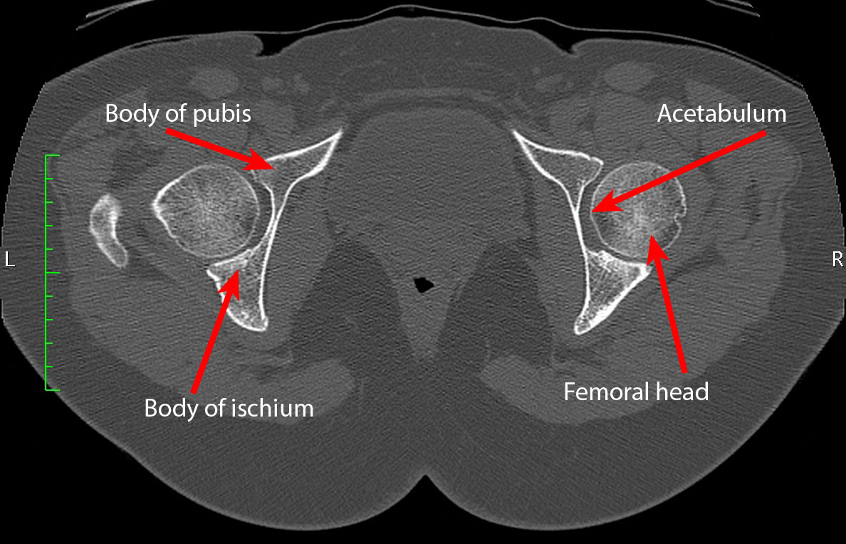
The hip is often considered the most difficult joint to arthroscope for several reasons 4 including the need for special equipment the local soft-tissue anatomy and the contour of the articulating surfaces of the femoral head and the acetabulum necessitating mechanical traction in order to obtain access to the joint. 25-7 Experienced hip arthroscopists have reported low. This CT imaging of the temporal bone was performed on a normal subject.
This CT imaging of the temporal bone was performed on a normal subject.
25-7 Experienced hip arthroscopists have reported low. 25-7 Experienced hip arthroscopists have reported low. This CT imaging of the temporal bone was performed on a normal subject. The hip is often considered the most difficult joint to arthroscope for several reasons 4 including the need for special equipment the local soft-tissue anatomy and the contour of the articulating surfaces of the femoral head and the acetabulum necessitating mechanical traction in order to obtain access to the joint.