Frontal Bone Skeletal Anatomy Next. Similarly the shapes of some muscles are very distinctive and the names such as orbicularis reflect the shape. The hard outer layer of bones is composed of compact bone tissue so-called due to its minimal gaps and spaces.
Initiated in 2017 by Dr. The cranium bones are called the ethmoid and frontal. A gomphosis fastened with bolts is the specialized fibrous joint that anchors the root of a tooth into its bony socket within the maxillary bone upper jaw or mandible bone lower jaw of the skull.
Similarly the shapes of some muscles are very distinctive and the names such as orbicularis reflect the shape.
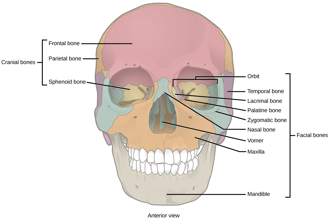
The hard outer layer of bones is composed of compact bone tissue so-called due to its minimal gaps and spaces. Frame of the body - the bones of the head and the trunk. The frontal bone forms the forehead the bony projections under the eyebrows and the superior part of each eyes orbits. The sphenoid bone is an unpaired bone of the neurocraniumIt is situated in the middle of the skull towards the front in front of the basilar part of the occipital boneThe sphenoid bone is one of the seven bones that articulate to form the orbitIts shape somewhat resembles that of a butterfly or bat with its wings extended.
