Frontal Bone Of Skull Anatomy. The right and left halves of the maxilla are irregularly shaped bones that fuse together in the middle of the skull below the nose in. The cranial roof consists of the frontal occipital and two parietal bones.
The fusion of these frontal bones forms a metopic suture which may be visible in some skull specimens. The maxilla is the bone that forms your upper jaw. A skull CT scan uses special X-ray equipment to generate a series of cross-sectional and three-dimensional images of the head and neck region1.
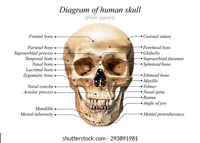
The entire skull is made up of 22 bones.
Sinuses are mucosa-lined airspaces within the bones of the face and skullEach opens into the anterior part of the corresponding middle nasal meatus of the nose through the frontonasal duct which traverses the anterior part of the labyrinth of the ethmoid. The frontal bone also forms the supraorbital margin of the orbit. The right and left halves of the maxilla are irregularly shaped bones that fuse together in the middle of the skull below the nose in. The name for the bone was derived from a deity of Greek mythology called Atlas.
